
Պատկեր:DTI-sagittal-fibers.jpg
Նախադիտման չափ՝ 643 × 600 պիքսել։ Այլ թույլտվությաններ: 257 × 240 պիքսել | 515 × 480 պիքսել | 1021 × 952 պիքսել.
Սկզբնական նիշք (1021 × 952 փիքսել, նիշքի չափը՝ 294 ԿԲ, MIME-տեսակը՝ image/jpeg)
|
Ամփոփում
| Նկարագրում |
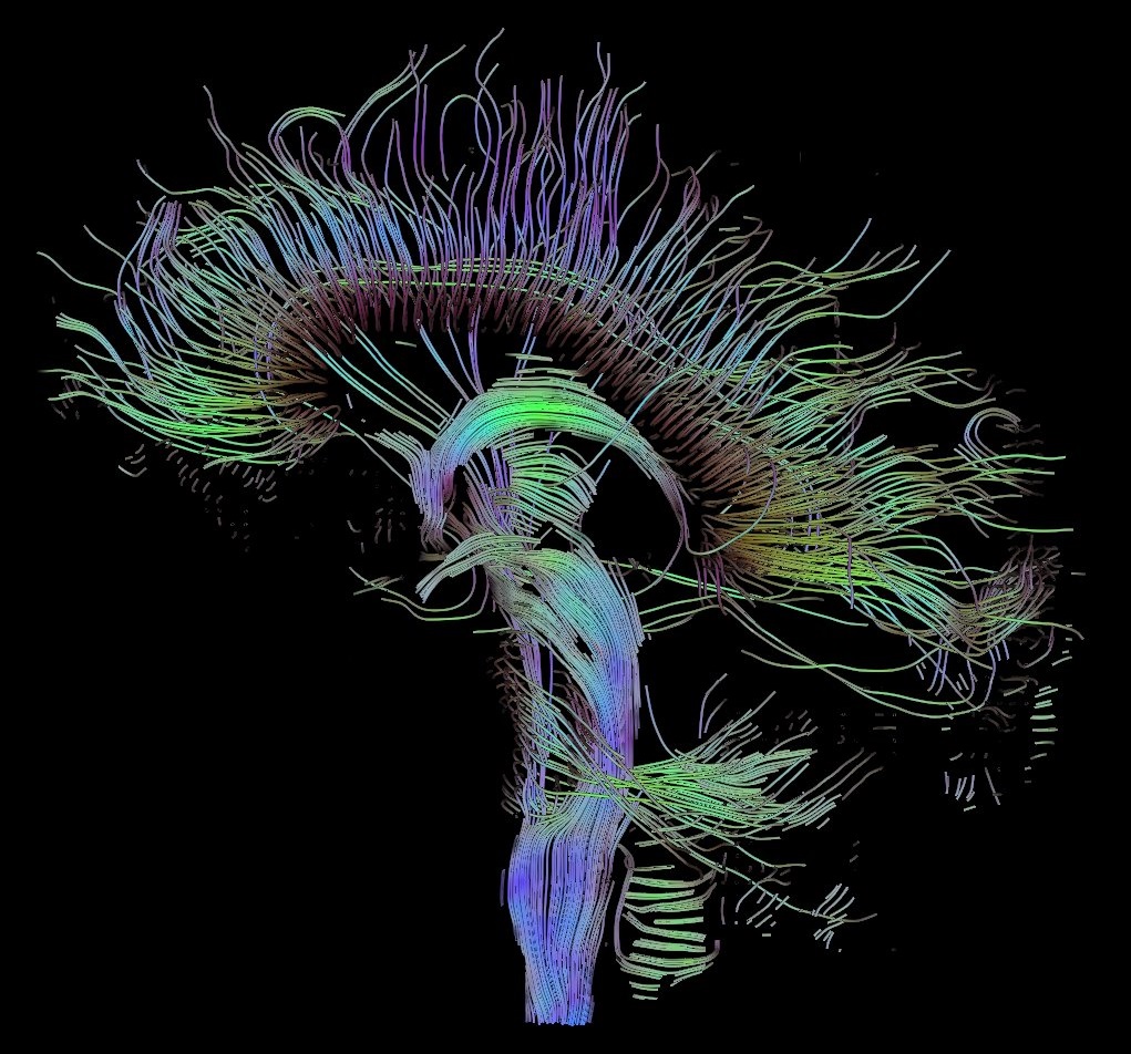
English: Visualization of a DTI measurement of a human brain. Depicted are reconstructed fiber tracts that run through the mid-sagittal plane. Especially prominent are the U-shaped fibers that connect the two hemispheres through the corpus callosum (the fibers come out of the image plane and consequently bend towards the top) and the fiber tracts that descend toward the spine (blue, within the image plane)
Français : Visualisation d'une mesure DTI d'un cerveau humain. Ce qui est représenté sont des faisceaux de fibres reconstruits qui traversent le plan demi-sagittal. On observe les fibres en U qui connectent les deux hémisphères à travers le corps calleux, qui sont particulièrement importantes (les fibres sortent du plan de l'image et par conséquent se courber vers le haut) ainsi que les faisceaux de fibres qui descendent vers la colonne vertébrale (bleu, dans le plan de l'image)
Deutsch: Traktographie-Verfahren rekonstruieren aus den Messdaten der Diffusions-Tensor-Bildgebung den anzunehmenden Verlauf größerer Nervenbahnen. Hier dargestellt sind die Ergebnisse für ein menschliches Gehirn; um die Übersichtlichkeit zu wahren, beschränkt sich die Abbildung auf Bahnen, die die Medianebene schneiden. Insbesondere sind dies die U-förmigen Faserbündel, die die beiden Hirnhälften verbinden (sie durchstoßen die Bildebene und sind nach oben gebogen) sowie die Faserbündel, die zum Rückenmark ziehen (blau dargestellt, liegen innerhalb der Bildebene) |
| Թվական | |
| Աղբյուր | Բեռնողի սեփական աշխատանք |
| Հեղինակ | Thomas Schultz |
| Իրավունքներ (Նիշքի վերաօգտագործումը) |
Rendering is own work, using a modified version of the BioTensor application developed at the University of Utah. The dataset is courtesy of Gordon Kindlmann at the Scientific Computing and Imaging Institute, University of Utah, and Andrew Alexander, W.M. Keck Laboratory for Functional Brain Imaging and Behaviour, University of Wisconsin, Madison. It is publicly available from [1] |
Արտոնագրում
Ես, այս աշխատանքի հեղինակային իրավունքների տերը, ներկա հրատարակում եմ սրա հետևյալ լիցենզիաների պայմաններով

|
Այս վավերագրման պատճենահանման, տարածման և/կամ ձևափոխման թույլտվություն կամ լիցենզիայի GNU FDL պայմաններով 1.2 մեկնակերպի կամ ավելի ուշ, հրատարակված Ազատ ծրագրային ապահովության հիմնադրամում, առանց անփոփոխելի հատվածների, առանց բնագիրների, որոնք տեղադրված են առաջին և վերջին շապիկներում: Լիցենզիայի պատճենը գտնվում է GNU Free Documentation License հատվածում: |
| Այս նիշքը հասանելի է Creative Commons Attribution-Share Alike 3.0 Unported արտոնագրի ներքո: | ||
| ||
| Այս արտոնգրության հատկանիշը ավելացված է տվյալ նիշքին որպես GFDL արտոնագրի բարեփոխում: |
This file is licensed under the Creative Commons Attribution-Share Alike 2.5 Generic, 2.0 Generic and 1.0 Generic license.
- Դուք ազատ եք՝
- կիսվել ստեղծագործությամբ – պատճենել, տարածել և փոխանցել այս աշխատանքը։
- վերափոխել – ադապտացնել աշխատանքը
- Պահպանելով հետևյալ պայմանները'
- հղում – Դուք պետք է նշեք հեղինակի (իրավատիրոջ) հղումը:
- համանման տարածում – Եթե դուք ձևափոխում եք, փոխակերպում, կամ այս աշխատանքի հիման վրա ստեղծում եք նոր աշխատանք, ապա ձեր ստեղծածը կարող է տարածվել միայն նույն կամ համարժեք թույլատրագրով։
Կարող եք ընտրել այս թույլատրագրերից ցանկացածը։
Նիշքի պատմություն
Մատնահարեք օրվան/ժամին՝ նիշքի այդ պահին տեսքը դիտելու համար։
| Օր/Ժամ | Մանրապատկեր | Օբյեկտի չափը | Մասնակից | Մեկնաբանություն | |
|---|---|---|---|---|---|
| ընթացիկ | 10:42, 13 Հոկտեմբերի 2017 | | 1021 × 952 (294 ԿԲ) | Mikael Häggström | Minor crop of black areas at the top and bottom |
| 16:22, 22 Սեպտեմբերի 2006 |  | 1021 × 1125 (203 ԿԲ) | Thomas Schultz | {{Information |Description=Visualization of a DTI measurement of a human brain. Depicted are reconstructed fiber tracts that run through the mid-sagittal plane. Especially prominent are the U-shaped fibers that connect the two hemispheres through the corp |
Նիշքի օգտագործում
Հետևյալ 2 էջերը հղվում են այս նիշքին՝
Նիշքի համընդհանուր օգտագործում
Հետևյալ այլ վիքիները օգտագործում են այս նիշքը՝
- Օգտագործումը af.wikipedia.org կայքում
- Օգտագործումը ar.wikipedia.org կայքում
- Օգտագործումը az.wikiquote.org կայքում
- Օգտագործումը bn.wikipedia.org կայքում
- Օգտագործումը cs.wikipedia.org կայքում
- Օգտագործումը de.wikipedia.org կայքում
- Autismus
- Computergrafik
- Bipolare Störung
- Portal:Informatik/Exzellente Artikel
- Portal:Geist und Gehirn/Artikel des Monats
- Diffusions-Tensor-Bildgebung
- Wikipedia:Kandidaten für exzellente Bilder/Archiv2006/17
- Datei:DTI-sagittal-fibers.jpg
- Wikipedia:Exzellente Bilder/Naturwissenschaften
- Portal:Physik/Artikel des Monats 2024-03
- Wikipedia:Exzellente Bilder/Kleine Bilder
- Օգտագործումը en.wikipedia.org կայքում
- Neurolinguistics
- Tractography
- Portal:Medicine
- User talk:Spikebrennan
- User:Spikebrennan
- Diffusion MRI
- Wikipedia:WikiProject Neuroscience
- Portal:Psychology/Selected article
- Wikipedia:Featured pictures/Sciences/Biology
- Portal:Psychology/Selected article/7
- Wikipedia:Featured pictures thumbs/08
- Wikipedia:Featured picture candidates/DTI-sagittal-fibers.jpg
- Wikipedia:Wikipedia Signpost/2007-11-05/Features and admins
- Wikipedia:Featured picture candidates/November-2007
- Wikipedia:Picture of the day/March 2008
- Connectome
- Template:POTD/2008-03-10
- User talk:Thomas Schultz
- Wikipedia:Wikipedia Signpost/2007-11-05/SPV
- Biological data visualization
- Wikipedia:WikiProject Medicine/Recognized content
- Wikipedia:WikiProject Molecular Biology/Biophysics
- User:Wouterstomp/test
- Wikipedia:WikiProject Anatomy/Resources
- Wikipedia:WikiProject Anatomy/Recognized content
- Wikipedia talk:WikiProject Anatomy/Archive 9
- Portal:Medicine/Recognized content
- User talk:Rhododendrites/Reconsidering FPC on the English Wikipedia
- User:Hydrogenkitsch
- Wikipedia:Wikipedia Signpost/Single/2007-11-05
- Օգտագործումը en.wikibooks.org կայքում
Տեսնել այս նիշքի ավելի համընդհանուր օգտագործումը: