
Պատկեր:Fundus of patient with retinitis pigmentosa, mid stage.jpg
Նախադիտման չափ՝ 699 × 599 պիքսել։ Այլ թույլտվությաններ: 280 × 240 պիքսել | 560 × 480 պիքսել | 871 × 747 պիքսել.
Սկզբնական նիշք (871 × 747 փիքսել, նիշքի չափը՝ 107 ԿԲ, MIME-տեսակը՝ image/jpeg)
| Նկարագրում |
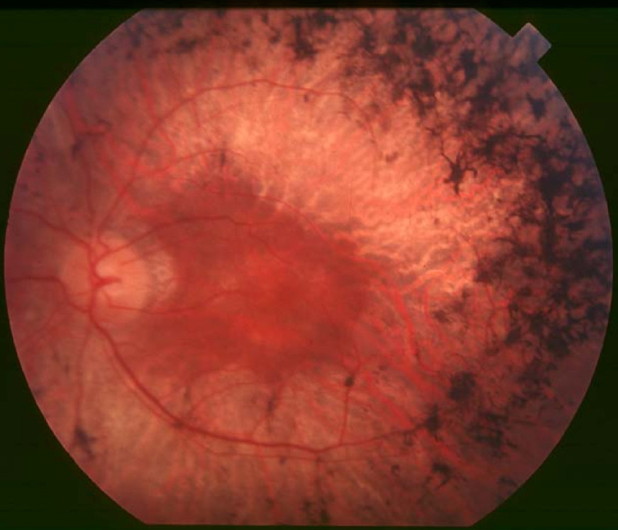
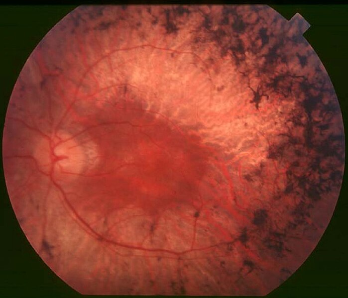
English: Figure 2. Fundus of patient with retinitis pigmentosa, mid stage (Bone spicule-shaped pigment deposits are present in the mid periphery along with retinal atrophy, while the macula is preserved although with a peripheral ring of depigmentation. Retinal vessels are attenuated.) Hamel Orphanet Journal of Rare Diseases 2006 1:40 doi:10.1186/1750-1172-1-40 |
| Թվական | |
| Աղբյուր | Retinitis pigmentosa by Christian Hamel |
| Հեղինակ | Christian Hamel |
| Իրավունքներ (Նիշքի վերաօգտագործումը) |
© 2006 Hamel; licensee BioMed Central Ltd. This is an Open Access article distributed under the terms of the Creative Commons Attribution License (https://creativecommons.org/licenses/by/2.0), which permits unrestricted use, distribution, and reproduction in any medium, provided the original work is properly cited. |
This file is licensed under the Creative Commons Attribution 2.0 Generic license.
- Դուք ազատ եք՝
- կիսվել ստեղծագործությամբ – պատճենել, տարածել և փոխանցել այս աշխատանքը։
- վերափոխել – ադապտացնել աշխատանքը
- Պահպանելով հետևյալ պայմանները'
- հղում – Դուք պետք է նշեք հեղինակի (իրավատիրոջ) հղումը:
Նիշքի պատմություն
Մատնահարեք օրվան/ժամին՝ նիշքի այդ պահին տեսքը դիտելու համար։
| Օր/Ժամ | Մանրապատկեր | Օբյեկտի չափը | Մասնակից | Մեկնաբանություն | |
|---|---|---|---|---|---|
| ընթացիկ | 10:17, 2 Դեկտեմբերի 2017 | | 871 × 747 (107 ԿԲ) | Doc James | Cropped 27 % horizontally and 7 % vertically using CropTool with precise mode. |
| 13:52, 22 Սեպտեմբերի 2009 |  | 1200 × 799 (126 ԿԲ) | CopperKettle | {{Information |Description={{en|1=Figure 2. Fundus of patient with retinitis pigmentosa, mid stage (Bone spicule-shaped pigment deposits are present in the mid periphery along with retinal atrophy, while the macula is preserved although with a peripheral |
Նիշքի օգտագործում
Հետևյալ էջը հղվում է այս նիշքին՝
Նիշքի համընդհանուր օգտագործում
Հետևյալ այլ վիքիները օգտագործում են այս նիշքը՝
- Օգտագործումը ar.wikipedia.org կայքում
- Օգտագործումը bs.wikipedia.org կայքում
- Օգտագործումը ca.wikipedia.org կայքում
- Օգտագործումը da.wikipedia.org կայքում
- Օգտագործումը en.wikipedia.org կայքում
- Օգտագործումը en.wikiversity.org կայքում
- Օգտագործումը es.wikipedia.org կայքում
- Օգտագործումը eu.wikipedia.org կայքում
- Օգտագործումը fa.wikipedia.org կայքում
- Օգտագործումը fi.wikipedia.org կայքում
- Օգտագործումը fr.wikipedia.org կայքում
- Օգտագործումը he.wikipedia.org կայքում
- Օգտագործումը it.wikipedia.org կայքում
- Օգտագործումը ko.wikipedia.org կայքում
- Օգտագործումը la.wikipedia.org կայքում
- Օգտագործումը or.wikipedia.org կայքում
- Օգտագործումը outreach.wikimedia.org կայքում
- Օգտագործումը pl.wikipedia.org կայքում
- Օգտագործումը pt.wikipedia.org կայքում
- Օգտագործումը ru.wikipedia.org կայքում
- Օգտագործումը sl.wikipedia.org կայքում
- Օգտագործումը sr.wikipedia.org կայքում
- Օգտագործումը sv.wikipedia.org կայքում
- Օգտագործումը th.wikipedia.org կայքում
- Օգտագործումը tr.wikipedia.org կայքում
- Օգտագործումը tt.wikipedia.org կայքում
- Օգտագործումը uk.wikipedia.org կայքում
Տեսնել այս նիշքի ավելի համընդհանուր օգտագործումը: