
Պատկեր:Virus Replication.svg

Սկզբնական նիշք (SVG-նիշք, անվանապես 462 × 426 փիքսել, նիշքի չափը՝ 205 ԿԲ)
Ամփոփում
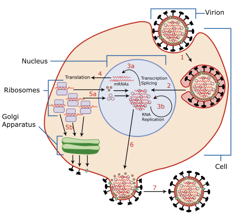
| Նկարագրում | A diagram of influenza viral cell invasion and replication. |
| Թվական | |
| Աղբյուր | Redrawn from w:Image:Virusreplication.png using Adobe Illustrator. |
| Հեղինակ | User:YK Times |
| Այլ մեկնակերպներ |
 |


|
Այս SVG ֆայլ օգտագործում է զետեղված տեքստ, որը կարելի է հեշտ թարգմանել ձեր լեզվի վրա using [toolforge:svgtranslate/File:Virus_Replication.svglink this automated tool]: Իմացեք ավելին: You can also download it and translate it manually using a text editor. |
Description from Scheme of Influenza A virus replication (NCBI): "A virion attaches to the host cell membrane via HA and enters the cytoplasm by receptor-mediated endocytosis (STEP 1), thereby forming an endosome. A cellular trypsin-like enzyme cleaves HA into products HA1 and HA2 (not shown). HA2 promotes fusion of the virus envelope and the endosome membranes. A minor virus envelope protein M2 acts as a ion channel thereby making the inside of the virion more acidic. As a result, the major envelope protein M1 dissociates from the nucleocapsid and vRNPs are translocated into the nucleus (STEP 2) via interaction between NP and cellular transport machinery. In the nucleus, the viral polymerase complexes transcribe (STEP 3a) and replicate (STEP 3b) the vRNAs. Newly synthesized mRNAs migrate to cytoplasm (STEP 4) where they are translated. Posttranslational processing of HA, NA, and M2 includes transportation via Golgi apparatus to the cell membrane (STEP 5b). NP, M1, NS1 (nonstructural regulatory protein - not shown) and NEP (nuclear export protein, a minor virion component - not shown) move to the nucleus (STEP 5a) where they bind freshly synthesized copies of vRNAs. The newly formed nucleocapsids migrate into the cytoplasm in a NEP-dependent process and eventually interact via M1 with a region of the cell membrane where HA, NA and M2 have been inserted (STEP 6). Then the newly synthesized virions bud from infected cell (STEP 7). NA destroys the sialic acid moiety of cellular receptors, thereby releasing the progeny virions."
Արտոնագրում

|
Այս վավերագրման պատճենահանման, տարածման և/կամ ձևափոխման թույլտվություն կամ լիցենզիայի GNU FDL պայմաններով 1.2 մեկնակերպի կամ ավելի ուշ, հրատարակված Ազատ ծրագրային ապահովության հիմնադրամում, առանց անփոփոխելի հատվածների, առանց բնագիրների, որոնք տեղադրված են առաջին և վերջին շապիկներում: Լիցենզիայի պատճենը գտնվում է GNU Free Documentation License հատվածում: |
| Այս նիշքը հասանելի է Creative Commons Attribution-Share Alike 3.0 Unported արտոնագրի ներքո: | ||
| ||
| Այս արտոնգրության հատկանիշը ավելացված է տվյալ նիշքին որպես GFDL արտոնագրի բարեփոխում: |
- Դուք ազատ եք՝
- կիսվել ստեղծագործությամբ – պատճենել, տարածել և փոխանցել այս աշխատանքը։
- վերափոխել – ադապտացնել աշխատանքը
- Պահպանելով հետևյալ պայմանները'
- հղում – Դուք պետք է նշեք հեղինակի (իրավատիրոջ) հղումը:
- համանման տարածում – Եթե դուք ձևափոխում եք, փոխակերպում, կամ այս աշխատանքի հիման վրա ստեղծում եք նոր աշխատանք, ապա ձեր ստեղծածը կարող է տարածվել միայն նույն կամ համարժեք թույլատրագրով։
| Annotations | This image is annotated: View the annotations at Commons |
Նիշքի պատմություն
Մատնահարեք օրվան/ժամին՝ նիշքի այդ պահին տեսքը դիտելու համար։
| Օր/Ժամ | Մանրապատկեր | Օբյեկտի չափը | Մասնակից | Մեկնաբանություն | |
|---|---|---|---|---|---|
| ընթացիկ | 02:54, 6 Մարտի 2007 | | 462 × 426 (205 ԿԲ) | YK Times | {{Information |Description=A diagram of influenza viral cell invasion and replication. |Source=Redrawn from w:Image:Virusreplication.png using Adobe Illustrator. |Date=March 5, 2007 |Author= User:YK Times |Permission= |other_versions=[[:w:Image:V |
Նիշքի օգտագործում
Հետևյալ էջը հղվում է այս նիշքին՝
Նիշքի համընդհանուր օգտագործում
Հետևյալ այլ վիքիները օգտագործում են այս նիշքը՝
- Օգտագործումը bg.wikipedia.org կայքում
- Օգտագործումը bn.wikipedia.org կայքում
- Օգտագործումը br.wikipedia.org կայքում
- Օգտագործումը ca.wikipedia.org կայքում
- Օգտագործումը cs.wikipedia.org կայքում
- Օգտագործումը da.wikipedia.org կայքում
- Օգտագործումը de.wikipedia.org կայքում
- Օգտագործումը el.wikipedia.org կայքում
- Օգտագործումը en.wikipedia.org կայքում
- Orthomyxoviridae
- Amantadine
- Viral replication
- Viral life cycle
- Viral entry
- File:Virusreplication.png
- User:YK Times/Graphic Lab/examples
- Wikipedia:Graphics Lab/Images to improve/Archive/Mar 2007
- Viral shedding
- Template:Influenza virus life cycle
- Viral neuraminidase
- Tilapia tilapinevirus
- User:Zoe.gum/sandbox
- Wikipedia talk:WikiProject Viruses/Archive 4
- User:Anicm1/sandbox
- Օգտագործումը en.wikibooks.org կայքում
- Օգտագործումը es.wikipedia.org կայքում
- Օգտագործումը fa.wikipedia.org կայքում
- Օգտագործումը fr.wikipedia.org կայքում
- Օգտագործումը id.wikipedia.org կայքում
- Օգտագործումը it.wikipedia.org կայքում
- Օգտագործումը ja.wikipedia.org կայքում
- Օգտագործումը kk.wikipedia.org կայքում
- Օգտագործումը ko.wikipedia.org կայքում
Տեսնել այս նիշքի ավելի համընդհանուր օգտագործումը: